Cruciate disease is the most common orthopaedic condition we see as small animal orthopaedic surgeons. However, at times it can still cause confusion in general practice, especially those subtle, early cases. This blog explores early cruciate disease with tips for early diagnosis and when to refer cases.
What is cruciate disease?
Cruciate disease that has progressed to complete rupture of the ligament can be a very easy diagnosis to make. Diagnosing cases earlier can be more challenging. It is not uncommon that we are referred dogs with early cruciate disease which have not been diagnosed as such and therefore have not been treated appropriately.
It’s important to realise that we call it a disease for a reason. Dogs rarely suffer truly traumatic cranial cruciate ligament (anterior cruciate ligament or ACL in human speak) ruptures. If one of us ruptured our ACL it would usually be a sporting injury – football players and skiers being particularly prone. In dogs it is very different. Canine cruciate disease is a degenerative process. We don’t fully understand the factors that initiate this process, but we do know that degeneration of the ligament ultrastructure progresses hand-in-hand with synovial inflammation. Over time this process results in a weaker ligament which will then start to tear.
The weakened ligament may tear acutely but often it’s a very gradual process. In the early stages of the disease, prior to substantial tearing of the ligament, the stifle is stable and the clinical signs (symptoms) can be subtle. In these early cases owners will commonly report that their dog will show a transient lameness or stiffness of the affected limb when they rise from resting. This is particularly common if the rest was preceded by a period of exercise. The lameness may only last a few seconds and there is often no lameness at other times. As the disease progresses these episodes will become more frequent and more obvious.
How to recognise the early signs
So how do we recognise these early cases, if there is no cranial drawer or cranial tibial thrust? Well, the history described above is non-specific but is very suggestive. On clinical examination the affected stifle may be thickened medially (‘medial buttress’). There may also be effusion, recognised by loss of definition of the edges of the patellar tendon.
A consistent finding is discomfort when the stifle is fully extended, which stresses the degenerate ligament. Be sure to partially extend the hip when assessing this, or stifle extension will also stretch the hamstring muscles. This also results in a pain response and can be misleading to the diagnosis.
Osteoarthritis and underlying disease
It’s likely that you are going to choose to radiograph a dog with such signs, if they have not resolved after a period of rest and NSAIDs, and here lies the key message. If you identify effusion and osteophytes, which are the typical changes associated with osteoarthritis (OA) then do not assume that the diagnosis is simply OA. It’s actually very rare for dogs to get primary OA. In fact, OA in dogs is almost always secondary to another joint issue. Most dogs with elbow OA have underlying elbow dysplasia, and most dogs with hip OA have hip dysplasia. The vast majority of cases of stifle OA will be secondary to cruciate disease. Employing the usual OA management strategies is not likely to help these dogs, since these strategies will not address the underlying cruciate problem.
How to manage cruciate disease in dogs
So what should you do in such cases? If you are presented with a patient with the history described above and with OA changes in the stifle, I would suggest an early referral. Resist the temptation to reach for more NSAIDs and wait to see if things get better or worse. Cruciate disease is invariable a one-way process. The ligament will continue to degenerate/tear, but it can be months (sometimes a year or more) before the stifle becomes unstable and the diagnosis becomes obvious. Throughout this time the joint will become more and more degenerate (‘arthritic’) and the clinical signs are likely to be persistent.
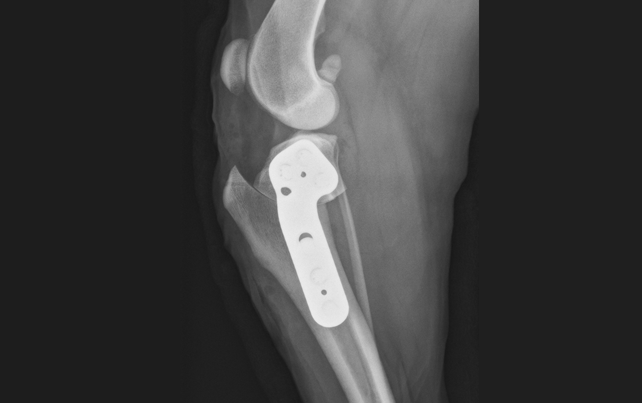
Surgery for cruciate disease in dogs
If confident in the diagnosis, then I will recommend TPLO surgery for early cruciate cases, even if the stifle is stable. Early TPLO can:
- Halt the progression of clinical signs
- Slow the degenerative process
- Reduce stresses on the ligament, which offers the opportunity to salvage what remains of the ligament and to maintain some intrinsic joint stability
- Reduces the risk of a meniscal tear
It is worth mentioning that TPLO is the surgery of choice at this stage. Other cruciate surgeries, such as TTA or lateral suture stabilisation, will not reliably achieve the same results. There are numerous studies now which clearly show that of all the cruciate surgeries, TPLO is the most reliable at providing stability and returning dogs to full function. Providing the best possible stability is particularly important for early cases where the intention is to salvage the ligament, otherwise the ligament is likely to continue to tear and cause inflammation.
Andy Moores
Andy Moores is one of the most qualified and experienced small animal orthopaedic surgeons in the UK. He has been a referral-only orthopaedic surgeon for over 20 years, a European Specialist for 18 years and an RCVS Specialist for 17 years. He has treated thousands of cats and dogs with orthopaedic problems during this time. This experience will be available to your clients and patients at The Moores Orthopaedic Clinic from May 2023. If you would like to discuss a case or make a referral then you can contact Andy directly via andy@mooresortho.com. You can subscribe to our newsletter and be informed of future blogs and articles at www.mooresortho.com